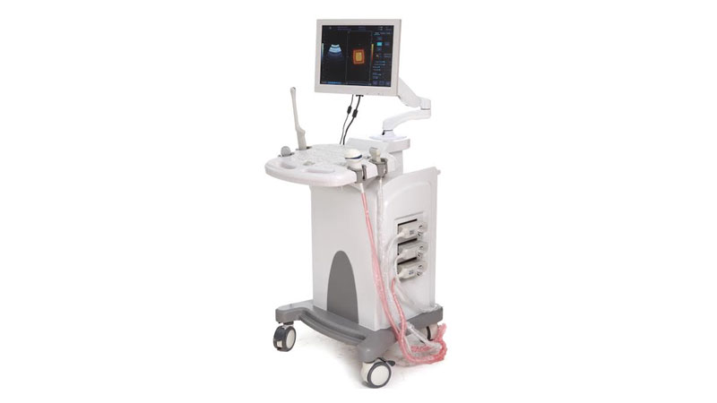
iLC Series Trolley Type Color Doppler

Technical Specifications :
- Imaging Technologies:
- High-precision digital continuous beam former
- Dynamic frequency fusion imaging
- High-precision delayed point-to-point dynamic receiving and focusing
- Superwide frequency imaging
- Adaptive image optimization
- Adaptive vascular imaging
- Adaptive Doppler imaging
- THI
- Applicable for the whole body, including but not limited to abdomen, Gyn., Obs., Cardiology, Vasuclar, Urology, Small organs, Mammary gland, paediatrics and neonatus, fetal cardiac imaging, puncture.
- Power :19.5V, 11.8A DC input
- Display : 17 / 19 / 21 inches medical grade monitor
- Probe Ports : 3 activated probe ports supporting convex, linear, phased array, endocavitary probes.
- Integrative network ultrasonic workstation. Available for DICOM.
- Scanning modes :B, M, CFM, PDI, PW, THI
- Image processing technologies
- Acoustic beam processing: full digital multi-beam former, real-time point-to-point dynamic receiving and focusing, continuous dynamic focusing, real-time dynamic variable aperture imaging, real-time dynamic acoustic beam apodization, dynamic filtering, dynamic frequency scanning.
- Image pretreatment
- Overall gain: 0~100 adjustable
- TGC:8 TGC sliders
- Gain control:B+M adjustable, CFM adjustable, PDI adjustable, PW adjustable
- Acoustic output: Low, middle, high adjustable
- Grey levels: grade 0 to 15 adjustable
- Digital channels: 32
- High-precision digital continuous beam former
- Edge enhancement 5 grades adjustable
- Frame average 6 grades adjustable
- Line average 3 grades adjustable
- Gamma correction grade 0 to 30 adjustable
- Contrast: 0~100
- Brightness: -50~+50
- Scanning parameters
- B mode
- Acoustical power: Low, middle, high adjustable
- B gains: 0-100
- Danymic range: 120dB
- M mode
- M acoustical power: Low, middle, high adjustable
- B gains: 0-100
- M scanning speed: Low, middle, high adjustable
- M sampling line visible & adjustable
- CFM color Doppler mode
- CFM gain: 0-100
- CFM wall filtering: 4 steps
- CFM baseline adjustment: 9 steps
- CFM frame average: 4 steps
- Maximum PRF:14kHz
- Minimum PRF:0.5kHz
- CDE Power Doppler mode
- CDE gain: 0-100
- PD frame average: 4 steps
- PD wall filtering: 4 steps
- Maximum PRF:14kHz
- Minimum PRF:0.5kHz
- PW Spectral Doppler mode
- PW gain:0-100
- PW dynamic range: 4 steps
- PW wall filtering: 4 steps
- PW color
- Digital wall filtering: 4 steps
- PW scanning speed: 4 steps (120,180,240,300)
- Sampling volume width: 1-15mm
- Angle correction
- Baseline adjustment: 9 steps
- Maximum PRF(speed):15kHz
- Minimum PRF(speed):1kHz
- Maximum detectable speed:21m/s(7.18m/s)
- Minimum detectable speed:0.05cm/s
- B mode
- Image display
- 256 gray scale
- Grey histogram display
- Image rotation : left / right, up / down, 90oC
- Depth : 3 to 24 cm (22 grades) depending on probes
- Emit focus : maximum 8 focus points (depending on probe types and depth)
- Dynamic range : ≥120dB (visible & adjustable)
- Real-time zoom : 6 times zoon in, zoom rate adjustable
- M mode speed : 3 grades adjustable
- Angle change : 3 angles (available for convex probe)
- Display TGC curve
- Video output : auto-select NTSC or PAL video format
- Screen display information
- Header information area: hospital log, hospital name, system date and time, patient information, TI/MI value, probe model and current mode.
- Menu adjustment area: image adjustment and optimization; pseudo colors, rotation and reverse; dynamic range, frame average, linear average, linear density, acoustic power of image real-time scanning; remark texts of current mode; body mark of current mode;basic measurement and applicable measurement items of current model under frozen condition.
- Parameter and measurement area: parameters of current image scanning; related measurement and calculation results when measuring.
- Image area: to display images under each mode and measurements and each texts, arrows.
- Caliper area: to display depth reference caliper, grey grade caliper, CFM or power's speed caliper; TGC curve displays or hides automatically according to parameter setup.
- Record area: to display current image and status messages being saved in video buffer in live condition; to display the saved total frames and status message of current frame under frozon condition.
- Menu status area: to display the status of related parameters in both live and frozon conditions.
- Acoustic output information: TI, MI, acoustic power(PH2、PL2 、PM2)。
- Measurement / Calculation
- B mode normal measurements: distance, circumrence (Ellipse & method of loci), area (Ellipse & method of loci), volume (Ellipse, method of loci and two-planes models), ratio and angle.
- M mode normal measurements: distance, time, slope and heart rate.
- PW mode normal measurements: time, heart rate, velocity, acceleration, resistance index.
- CFM、PDI mode normal measurements:distance, circumrence (Ellipse & method of loci), area (Ellipse & method of loci), volume (Ellipse, method of loci and two-planes models), ratio and angle.
- Gynecological measurement: uterus (uterus, cervix and endometria), ovarian volume and left & right follicle.
- Obstetric measurement: EDD table :GS, BPD, CRL, FL, YS, TAD, LV, OFD, NT, AC, HC, APAD, Cxlength。
- Cardialogy measurement: applicable measurements for aortic, aortic vaivem, left atrium, LV, mitral vaive, right atrium, right ventricle, circulatory system.
- Urological measurment: prostate volume, seminal vesicle size, transition zone volume, bladder volume, testicular volume, residual urine volume, renal volume.
- Superficial organs measurement: thyroid gland.
- Others: calculations of hip angles by drawing baselines, ridge line and apical line.
- Measurement Reports
- Obs., Gyn., Cardiac, Urological and other measurement reports
- Measurement results saved automatically and generates reports
- Marks
- ≥95 body marks
- Text marks
- Arrow marks
- Storage
- Image storage, video storage, cine loop(≥300 frames), disk storage≥160G.
- Peripheral Devices
- Recorder, USB removable storage device
- Digital, video printers
- Laser, jet printers
- S-Video、AV video output
- Puncture guide line
- Puncture guide functions available for all probes.
- Angle of puncture guide line is adjustable and predefined.
Winning Advantages :
- Powerful software packages on Gyn. Obs, small organ, cardiac, abodomen, uorology, MSK, vessels, fetal etc.
- Image Quality is better and can be compared with renowned Global Brands.
- 17 / 19 / 21 inches LCD monitor, Fashionable design, In-built battery
- Triplex / Duplex, 3D / 4D optional, Advanced signals processing technology
- Excellent measurement and calculation programs for General, Obstetrics, Gynecology, Urology, Small Parts, Cardiac, Skeletal & Muscles Vascular examinations especially on vessels, Blood
" Ultrasound Scanner Machines will be Sold & Purchased strictly as per the applicable Guidelines of PC & PNDT."


