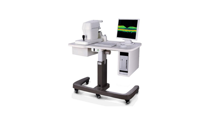
Optical Coherence Tomography - OCT

Technical Specifications :
| Tomographic Imaging | ||
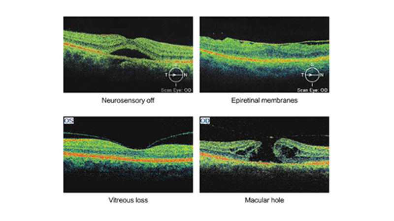
| Purpose | Cross Sectional Imaging of the Retina | |
| Signal Type | Photon scattering from tissue |
| Light Source | : | Super luminescent diode, 830nm |
| Optical Power | : | ≤ 0.7mW (on the cornea) |
| Axial Resolution | : | 5 - 8 um in tissue |
| Lateral Resolution | : | 15 um in tissue |
| Scanners | : | Galvanometer Mirror |
| Scan Mode | : | Line, Concentric ring, Repeat, Arbitrary-angle |
| Scan Rate | : | 400 A - scan/s |
| Acquisition Time | : | 1 sec |
| Scan Depth | : | 2mm in tissue |
| Fundus Imaging | ||
| Purpose | : | Fundus observation and real-time registration of OCT imaging |
| Signal Type | : | CCD imaging |
| Field Angle | : | 29°x 23° |
| Viewing Method | : | 15 inch color flat panel display |
| Illumination | : | LED |
| Internal Fixation | : | LED dot matrix |
| External Fixation | : | Adjustable blinking LED |
| Minimum Pupil Diameter | : | 3.5mm |
Winning Advantages:
- High Performance at a low price: Modular design increases flexibility, Reusability and Maintainability. Personalized design according to the customer's needs cab be provided.
- With the powerful software, it has clear, easy to use interface and support multiple language.
- Anterior Segment Analysis Template : Anterior segment with optional modules, able to observe and analyzer the anterior segment.


