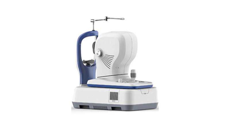
ivOC 30

Winning Advantages:
MACULA
- LSO:Equipped with LSO(Line Scanning Ophthalmoscopy)MOcean 30 provides simultaneously high quality fundus imaging, which is easy for physicians to localize the lesion.
- MACULA : Macula HD line : High definition OCT imaging reveals small lesions, OCT scan length can be switched between 6mm and 12mm.
- Macula Six-line Radial : Having a glimpse of the Retina with HD imaging and quick data analysis Software Analysis:Retinal Thickness Analysis, Ganglion Cell Analysis, High definition OCT imaging with 5 images averaging.
- Macula Cube : A point-by-point assessment of Retinal thickness with a 500x100 dense cube Software Analysis:Retinal thickness analysis,Progression analysis, 3D view,En-face analysis.
GLAUCOMA
- For comprehensive glaucoma analysis. MOcean 30/30 Plus offers two scan modes : glaucoma cube scan in macular area and glaucoma cube scan in disc area. Evenly distributed sampling rate with 200 x 200 A-scans provides reliable information for early glaucoma detection and management.
- Glaucoma(Macular): Software Analysis:Ganglion cell analysis, Progression analysis.
- Glaucoma (Disc) : Software Analysis:RNFL analysis, Cup-disk analysis,Calculation circle and circle scan tomogram, Progression analysis, OU comparative analysis.
- Informative Report : OU comparative analysis, Progression Analysis Report.
ANTERIOR SEGMENT
- Anterior HD Line:High - definition OCT imaging of the cornea enables localization of the Bowman's layer, the interface between corneal stroma and epithelium, Anterior Chamber Angle,Manual measurement is available.
- Anterior Six-line Radial:Anterior segment scanning through 6 radial lines of equal length can be used to measure the central corneal thickness, Software Analysis, Corneal Pachymetry,Manual Measurement.
PERMIUM FUNCTIONS
- En-face Analysis:En-face OCT provides ability to precisely localize lesions within specific sub retinal layers.
Choroid View
Choroid View
IS / OS-Ellipsoid View
Mid - Retina View
VRI View
3D En - face View - Network System : Remote Analysis System : Moptim provides a remote viewer software for displaying, enhancing, analyzing and saving digital images obtained with MOcean 30/30plus.
Remote Diagnosis System : Customer scan are reviewed remotely by specialists at Big Picture Eye Health for over 45 eye pathologies.
Software immediately generates a customer report, including educational content and specialist referral if needed.
Optional module, which is developed and operated by Big picture Eye Health, can be connected to MOcean 30 seamlessly.
